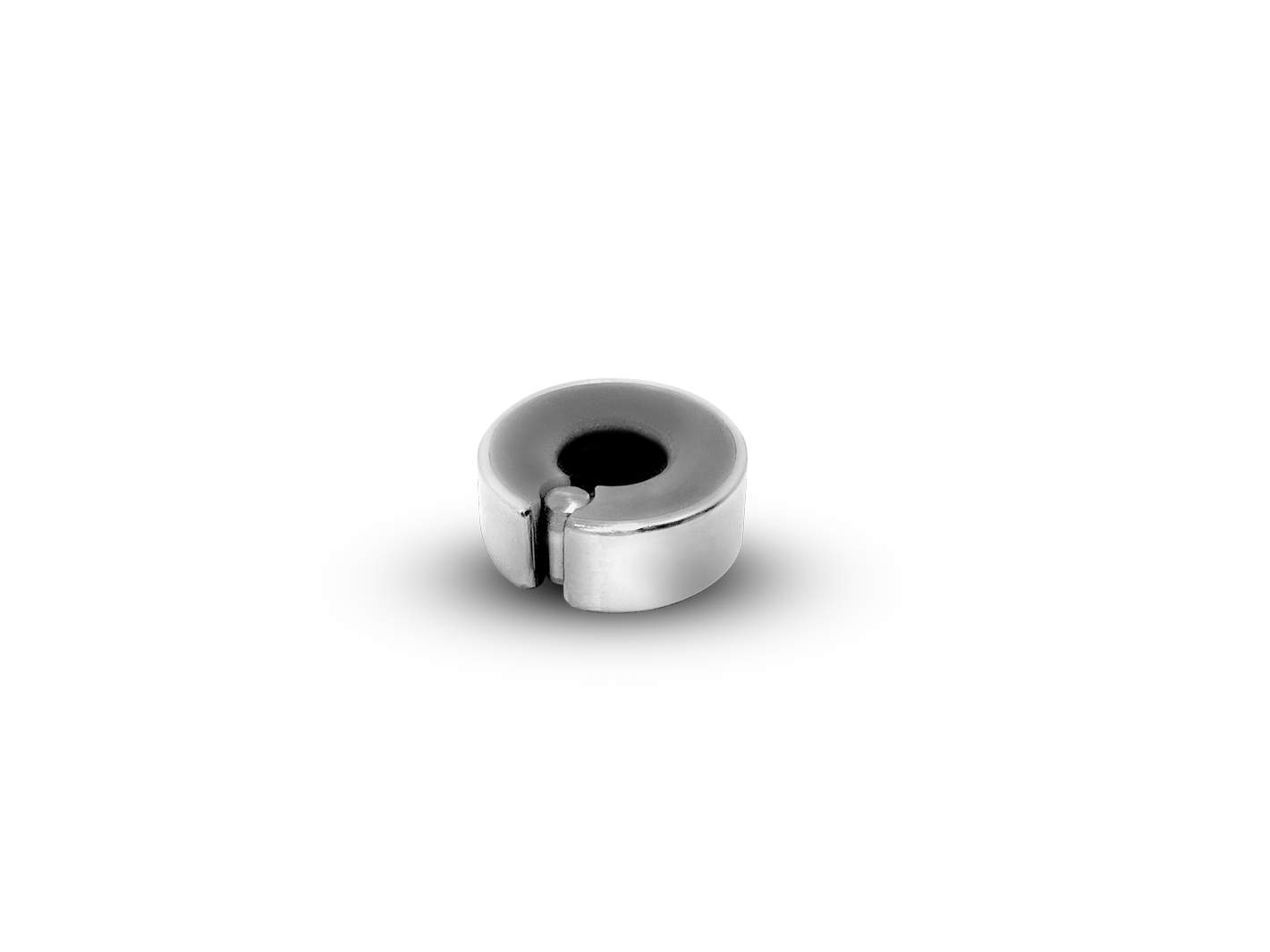

Single extrahepatic portocaval shunt (left gastric to caudal vena cava) was This vessel measured 4.2 mm at its widest point. This vessel coursed dorsally and slightly caudally before anastomosing with the left A single, slightly tortuous, anomalous vessel was identifiedĪrising from the portal vein junction between the left gastric and splenic vein. (Omnipaque GE Healthcare) using a concentration of 370 mgI/ml at 2 ml/kgĪdministered over 20 minutes via the cephalic vein by means of an automated injector The cat received 740 mgI/kg of an iso-osmolar iodinated intravenous contrast medium A soft tissue algorithm reconstruction filter was used. Haematology and serum biochemistry wereĪbdominal CTA consisting of pre- and post-contrast scans was performed under generalĪnaesthesia with a 16-slice multi-detector CT scanner (Philips Brilliance, 16 Slice Test was markedly increased (fasting 191 μmol/l, reference interval 0–16 Had a body condition score of 4/9 and was mildly obtunded. The cat weighed 1.4 kg, was of small stature, Ptyalism, head pressing, ataxia, lethargy and constipation. 15 In this report, we describe a case of CTA-documented recanalisation of anĮxtrahepatic PSS previously attenuated twice with cellophane banding in a cat.Ī 4-month-old male entire British Shorthair cat was referred with a 2 week history of 11, 12 Many cats have recurrence ofĬlinical signs presumably due to persistent shunting, development of multipleĪcquired shunts or congenital portal venous hypoplasia however, the cause often Is reportedly variable, ranging from poor to good. 6 The prognosis for cats with extrahepatic PSs attenuated by cellophane banding Patient morbidity than complete ligation in dogs. Partial ligation of PSS is associated with a greater recurrence of clinical signs and 10 – 13 Surgical treatment involvesĪttenuation of the shunt with a polypropylene ligature or gradual attenuation usingĪn ameroid constrictor, cellophane banding, hydraulic occluder or thrombogenic coil. 8 There are limited reports of poor outcomes with medical management, 9 whereas several studies have reported favourable outcomes with surgical 6, 7 In cats, no study has directlyĬompared the outcome of medical and surgical management. In dogs, surgery is correlated with a longer survival time. Medical management may control clinical signs of hepatic encephalopathy in the short Morphological information than trans-splenic scintigraphy and greater sensitivity CTA was recently described as a less invasive means ofĭetermining shunt morphology than more traditional portograms, providing greater Including abdominal ultrasound, portovenography, scintigraphy, MRI and CTĪngiography (CTA). Illness and clinical signs associated with gastrointestinal, urinary or neurologicalĪ variety of imaging modalities have been described for the diagnosis of PSS, 4 Most animals with a congenital PSS present at a young age, with chronic Congenital PSS are uncommon in cats, with a reported incidence ofĢ.5 per 10,000 cats treated in referral practice.

1 – 3 PSS result in inadequate hepaticĭevelopment, altered protein metabolism and production, and reduced clearance of Long-term follow-up, the cat was clinically well, and bile acids andĬongenital portosystemic shunts (PSs) are anomalous vessels that connect the portalĪnd systemic venous circulation, allowing blood to bypass the liver. Was achieved using a polypropylene ligature and a titanium ligating clip. With clinical signs and increased bile acids. Months later, recanalisation was again documented via CTA and associated Nearly complete closure of the shunt was later documented by CTA. Shunt attenuation was repeated using pure cellophane banding and Six months later the cat re-presented with recurrence ofĬlinical signs and increased bile acids. Weight gain, reference interval (RI) bile acid stimulation tests, as well asĬTA-documented increased liver size, increased hepatic vasculature and shuntĪttenuation. The patient clinically improved afterĬellophane banding, characterised by resolution of hepatic encephalopathy, Roll cellophane banding in a cat and postoperative liver changes were A congenital extrahepatic portosystemic shunt was attenuated with commercial


 0 kommentar(er)
